A negative MRI brain injury lawsuit often begins with a frustrating contradiction. The scan comes back "normal," but the headaches, memory gaps, and difficulty concentrating are anything but. For people recovering from collisions on I-91, I-84, or local Connecticut roads, hearing "your imaging looks fine" may feel like the injury itself is being dismissed.
Standard imaging was never designed to detect every type of brain damage. CT scans and conventional MRIs look for bleeding, fractures, and large structural abnormalities. When those results come back clear, it does not mean the brain is uninjured. It means the damage present may be invisible to those tools.
Key Takeaways for Negative MRI Brain Injury Lawsuits
- A "normal" MRI or CT scan does not rule out brain injury. These tools detect bleeding and structural damage but often miss microscopic injuries like diffuse axonal injury.
- Diffuse axonal injury, which involves shearing of microscopic brain fibers, is one of the most common forms of traumatic brain injury and frequently does not appear on standard scans.
- Advanced imaging tools like DTI and SWI may detect damage that conventional scans miss, and courts in some jurisdictions have considered this evidence in brain injury litigation.
- Connecticut law allows two years from the date of injury to file most personal injury claims under Connecticut General Statutes § 52-584, making early documentation of symptoms critical.
- Proving a brain injury without a positive scan relies on consistent symptom reporting, neuropsychological testing, and functional evidence rather than imaging alone.
Why Your MRI May Be "Normal" After a Brain Injury
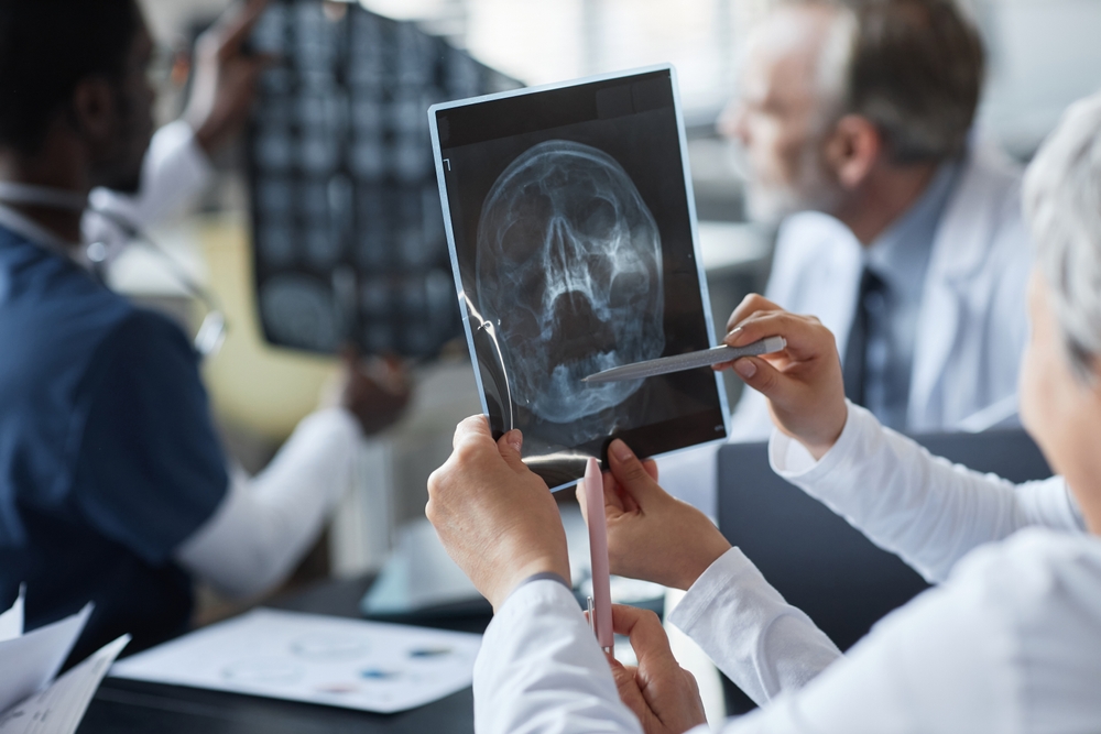
The experience of having a ‘normal’ MRI despite ongoing brain injury symptoms is one of the most frustrating parts of post-accident recovery. Understanding why this happens starts with knowing what these scans actually measure.
CT Scan vs. MRI Brain Injury Difference
A CT scan is often the first tool used in a Connecticut emergency room after a head injury. It is fast and effective at identifying acute bleeding and skull fractures, but it does not detect microscopic brain damage.
A standard MRI provides more detail about soft tissue and larger structural issues. However, it still misses the kind of damage that causes persistent cognitive symptoms after a concussion or mild TBI. The CT scan vs. MRI brain injury difference matters legally because adjusters often treat "normal" results from either scan as proof that no injury occurred.
Structural vs. Functional Imaging of Brain Injuries
Structural imaging looks at the physical architecture of the brain. Functional imaging looks at how the brain actually operates, including blood flow, neural connectivity, and white matter integrity.
This distinction between structural vs. functional imaging in a brain injury case is central to understanding why many legitimate injuries go undetected. A brain may look structurally intact while functioning abnormally in ways that affect memory, attention, and processing speed.
When a claim depends on proving injury, functional evidence often fills the gap that structural imaging leaves open.
Diffuse Axonal Injury: The Damage Standard Scans Miss
Diffuse axonal injury occurs when the brain shifts rapidly inside the skull, shearing microscopic nerve fibers called axons. This type of microscopic brain damage is frequent in car accidents, particularly rear-end collisions and high-speed impacts on highways like I-84 and Route 9.
Why Diffuse Axonal Injury Does Not Appear on a CT Scan
The National Institutes of Health recognizes diffuse axonal injury as a significant form of TBI. The damage occurs at the cellular level, involving individual nerve fibers too small for CT or standard MRI to detect. Diffuse axonal injury not being visible on a CT scan is not unusual. It is a well-documented limitation of emergency imaging.
When these axons are sheared or stretched, communication between brain regions is disrupted. The resulting symptoms, including difficulty concentrating, short-term memory problems, fatigue, and slowed processing, are real and measurable through clinical testing even when no scan shows visible damage.
From a legal perspective, this gap between "normal" imaging and persistent functional symptoms is where many brain injury claims are won or lost.
Proving TBI Without a Positive Scan in Connecticut
Proving TBI without a positive scan is one of the most challenging aspects of brain injury litigation. Insurance adjusters often point to clean imaging as evidence that no serious injury exists. Building a claim requires shifting the focus from structure to function.
Neuropsychological Testing as Core Evidence
Neuropsychological testing involves standardized assessments that measure cognitive function, including memory, attention, processing speed, language, and executive function. Unlike imaging, these tests measure how the brain performs rather than how it looks.
A neuropsychologist compares results against expected baselines. When scores fall below expected ranges in patterns consistent with brain injury, the testing provides objective, measurable evidence that supports the claim.
Building a Claim Through Consistent Documentation
Beyond neuropsychological testing, several types of evidence help prove a brain injury when scans are negative. Each type adds a layer that makes the claim harder to dismiss.
The following forms of evidence often strengthen a brain injury normal scan lawsuit in Connecticut:
- Consistent symptom reporting: Medical records showing the same cognitive complaints across multiple visits and providers establish a reliable pattern.
- Functional capacity evaluations: Formal assessments of a person's ability to perform work tasks and daily activities provide measurable data about limitations.
- Employment records: Documentation of reduced hours, job performance issues, or inability to return to work connects the injury to economic loss.
- Statements from family and coworkers: People who interact with the injured person regularly may observe changes in memory, behavior, or personality that medical records alone do not capture.
- Treatment consistency: A documented history of following medical recommendations strengthens credibility and counters arguments that symptoms are exaggerated.
Together, these forms of evidence create a picture that imaging alone cannot provide. A claim built on layered, consistent documentation is far more difficult for an insurer to reduce to a single "normal" scan result.
Advanced Imaging: DTI and SWI Scans in Brain Injury Cases
Two advanced imaging techniques may detect types of brain damage that conventional scans miss entirely. Their role in litigation is growing, though admissibility varies by jurisdiction and case.
DTI Scan Brain Injury Evidence
DTI, or diffusion tensor imaging, measures water molecule movement along white matter tracts in the brain. When axons are damaged, these movement patterns change. DTI scans provide visual maps of white matter integrity that may help a judge or jury understand injury that a standard MRI would never reveal.
DTI has been used in litigation to support claims involving diffuse axonal injury. The imaging adds a visual component to what might otherwise rely entirely on clinical testing and symptom reporting.
SWI MRI Brain Injury Detection
SWI, or susceptibility-weighted imaging, is an MRI technique designed to detect very small areas of bleeding called microbleeds. Standard MRI sequences often miss these.
SWI MRI brain injury detection may identify hemorrhagic evidence of injury that other scans overlook, particularly in cases involving rapid deceleration forces common in Connecticut highway collisions.
DTI Scan Admissibility in Connecticut Courts
DTI scan admissibility is not automatic. Connecticut courts evaluate scientific evidence under standards that consider whether the methodology is reliable and generally accepted in the relevant scientific community. Courts may require testimony from qualified professionals explaining the methodology and its application to the specific case.
As advanced imaging becomes more widely studied, its role in brain injury litigation continues to develop. A traumatic brain injury lawyer who is familiar with these evidentiary issues may understand how to present this evidence effectively within Connecticut's legal framework.
What Standard Scans Show vs. What They Miss
Imaging limitations help explain why a "normal" result does not end the medical or legal story.
| Imaging Type | What It Detects | What It Often Misses |
| CT Scan | Bleeding, skull fractures | Diffuse axonal injury, microscopic damage |
| Standard MRI | Larger structural abnormalities | Functional impairment, subtle shearing |
| DTI | White matter tract disruption | Not routinely used in ER settings |
| SWI | Microbleeds | May still miss functional deficits |
No single tool captures the full picture. A comprehensive approach to proving a brain injury often involves combining imaging with clinical testing and documented functional limitations.
How Insurance Companies Use "Normal" Scans to Reduce Claims
When an adjuster reviews a brain injury claim with normal CT and MRI results, the reasoning follows a predictable pattern: if the scan is normal, the injury must be minor. This logic has significant gaps, but it drives claim evaluations every day.
Common Adjuster Arguments Against Normal-Scan Claims
The following positions commonly appear in insurance evaluations of brain injury claims with negative imaging:
- "The scan is normal, so the injury is not serious." This treats imaging as the sole measure of injury, ignoring functional evidence and clinical testing.
- "Symptoms are subjective and unverifiable." This dismisses patient-reported symptoms without considering neuropsychological results or documented patterns.
- "Pre-existing conditions explain the symptoms." This shifts causation away from the accident, even when symptoms appeared or worsened after the incident.
- "Treatment gaps suggest the injury resolved." Periods without medical visits are framed as evidence of recovery rather than as barriers to access.
Recognizing these arguments helps explain why thorough documentation matters. Each potential challenge is best addressed before it becomes a reason to reduce or deny a claim.
Why Functional Evidence Changes the Equation
A claim that relies solely on imaging is vulnerable when that imaging is negative. A claim built on neuropsychological testing, consistent symptom documentation, and employment records shifts the focus from what the scan shows to what the person experiences.
TBI symptoms may persist even when standard imaging appears normal. This supports the position that a negative scan does not equal a negative outcome.
Building a Negative MRI Brain Injury Lawsuit in Connecticut
Filing a negative MRI brain injury lawsuit in Connecticut requires a strategic approach to evidence. Connecticut's two-year statute of limitations under § 52-584 applies to brain injury claims, making early documentation essential.
Several steps may help strengthen a brain injury claim when imaging is negative:
- Seek neuropsychological testing early. Formal cognitive assessments document impairment close to the time of injury.
- Report symptoms at every medical visit. Consistent reporting across multiple providers creates a documented pattern supporting the diagnosis.
- Keep a daily symptom journal. Recording headaches, memory problems, and mood changes provides a personal record that complements medical documentation.
- Request advanced imaging when appropriate. DTI or SWI scans may reveal damage that standard imaging missed.
- Preserve employment and financial records. Documentation of reduced work capacity ties the injury to measurable economic impact.
The strength of these cases comes from consistency and thoroughness, not from any one test or scan result.
FAQ for Negative MRI Brain Injury Lawsuits
Is it possible to have brain damage with a normal MRI?
What is the difference between a CT scan and an MRI for brain injuries?
A CT scan is faster and identifies acute bleeding and skull fractures. An MRI provides more detail about soft tissue and structural abnormalities. Neither tool reliably detects diffuse axonal injury or other microscopic damage that may cause persistent cognitive symptoms.
How is a brain injury proven in court without scan evidence?
Neuropsychological testing, consistent medical documentation, functional capacity evaluations, and testimony from family or coworkers may all serve as evidence. Courts evaluate the totality of evidence, not just imaging results.
Are DTI scans accepted as evidence in Connecticut courts?
DTI evidence may be admitted when the methodology is shown to be reliable and relevant to the specific case. Admissibility often depends on testimony from qualified professionals and the standards applied by the presiding judge.
How long do I have to file a brain injury lawsuit in Connecticut?
Connecticut law generally allows two years from the date of injury to file a personal injury claim. This deadline applies regardless of whether imaging shows visible damage. Consulting with an attorney early helps preserve options and evidence.
The Scan Does Not Tell the Whole Story
A normal MRI result does not define the boundaries of a brain injury claim. At The Flood Law Firm, our attorneys regularly handle complex brain injury cases that are referred by other Connecticut lawyers, including cases where standard imaging shows no visible damage.
We understand how to build claims around functional evidence, advanced imaging, and thorough documentation.
If you are living with cognitive symptoms that a "normal" scan does not explain, our team is ready to listen. Contact our Middletown office at (860) 346-2695 for a free consultation. We take calls in English and Spanish, and there are no upfront fees.